The photoacoustic effect is a phenomenon in which ultrasound waves are generated in biological tissues exposed to laser pulses. Detecting these ultrasound waves then allows biomedical imaging. Photoacoustic imaging can be useful, e.g., for cancer diagnostics, because using sound instead of light as a signal can improve the imaging resolution of deep tissue sites. However, the method can be hampered by ambient noise introduced by substances such as hemoglobin or melanin that are present in the body. Using targeted probes that are activated by, e.g., hypoxia, which is common in tumors, can improve the specificity of imaging.
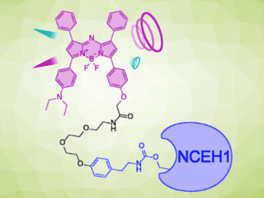
Janggun Jo, University of Michigan, Ann Arbor, USA, Jae Won Chang, Emory University, Atlanta, GA, USA, and colleagues have developed a photoacoustic imaging probe with dual-factor activation (activated form pictured). The probe is activated by hypoxia and also selectively and covalently binds to neutral cholesterol ester hydrolase 1 (NCEH1), a protein that is highly expressed and activated in several aggressive cancers. It is based on an aza-BODIPY (BODIPY = boron-dipyrromethene) dye and features an N-oxide group that is reduced to the corresponding amine under hypoxic conditions, as well as a linker for binding to NCEH1.
The probe showed absorbance and emission wavelengths that fall within the desired range for practical imaging, and the necessary stability in biological buffer solutions. In-vivo experiments showed promising results with strong signals and high specificity. The probe successfully visualized aggressive prostate cancer tumors in a mouse model.
- Development of a Dual Factor Activatable Covalent Targeted Photoacoustic Imaging Probe for Tumor Imaging,
Jiho Song, Tianqu Zhai, Heung Sik Hahm, Yuancheng Li, Hui Mao, Xueding Wang, Janggun Jo, Jae Won Chang,
Angew. Chem. Int. Ed. 2024.
https://doi.org/10.1002/anie.202410645
![]()