|
|
Entry 1
|
Entry 2 |
|
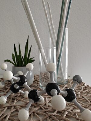
“Hexyllithium”
Photo by Ricarda Schmid
|
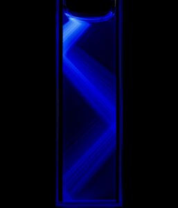
“Reflections”
The picture presents a total internal reflection of light passing through a solution of fluorescent dye
Photo by Borys Osmialowski
|
|
Entry 3
|
Entry 4 |
|
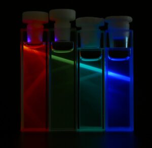
“A Colorful World”
Cuvettes with solutions containing different fluorophores
Photo by Borys Osmialowski
|
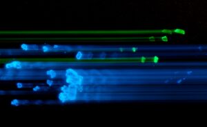
“Almost Collision”
The intentional camera movement results in a picture of moving crystals that, for some reasons, did not collide
Photo by Borys Osmialowski
|
|
Entry 5
|
Entry 6 |
|
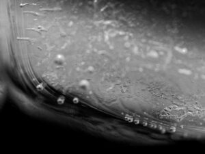
“Bubbles Under Ice”
The black and white picture shows the bubbles of air going up the ice cube and creating grooves in the ice
Photo by Borys Osmialowski
|
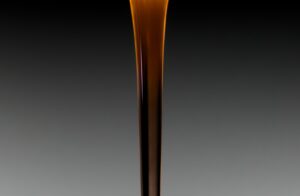
“Liquid Column”
A solution of fluorescent dye during flash chromatography
Photo by Borys Osmialowski
|
|
Entry 7
|
Entry 8 |
|
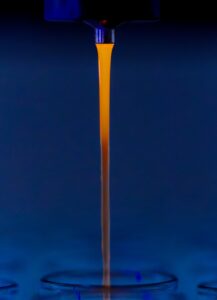
“Liquid column”
A solution of fluorescent dye during flash chromatography
Photo by Borys Osmialowski
|
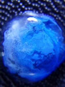
“A Microcosmic Blue Planet”
Macrophotography capturing the reaction of sodium hydroxide with copper sulfate, producing copper hydroxide as a blue precipitate in a drop of water.
Photo by Michele Rizzello
|
|
Entry 9
|
Entry 10
|
|
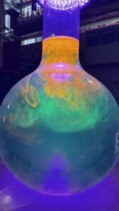
“Aurora Borealis in a Beaker”
Different UV-active solutions in a beaker
Photo by Tobias Kubo
|
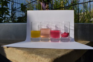
“Home-Made pH Indicator I”
A home-made pH indicator from rose petals. Using plants or products found at home for experiments can motivate students and open their mind to the world of chemistry (description of experiment)
Photo by Anya Slomba
|
|
Entry 11
|
Entry 12 |
|
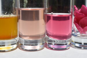
“Home-Made pH Indicator II”
A home-made pH indicator from rose petals. Using plants or products found at home for experiments can motivate students and open their mind to the world of chemistry (description of experiment)
Photo by Anya Slomba
|
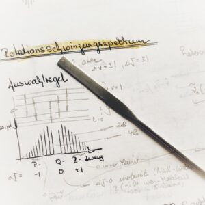
“Notes and Spatula”
Photo by Myriam Wójcik
|
|
Entry 13
|
Entry 14 |
|
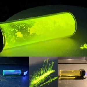
“Dipodal Fluorophore”
A hydrazone in its solid state and in solution, when exposed to UV and white light.
Photo by Kirk Wilson
|
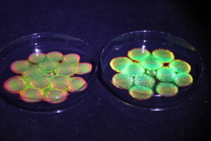
“Structure Color Colony of Microorganism”
The marine microorganism Cellulophaga sp. is known for its structurally colored colonies on agar. The iridescent color varies depending on the angle of illumination.
Photo by Mikiko Tsudome
|
|
|
Entry 15
|
Entry 16 |
|
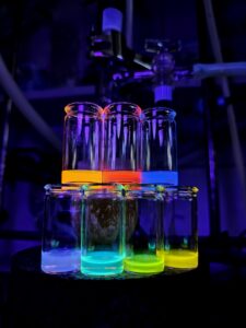
“Science Behind the Rainbow”
The rainbow world of fluorescence colors obtained in everyday work on scientific research
Photo by Maciej Majdecki
|
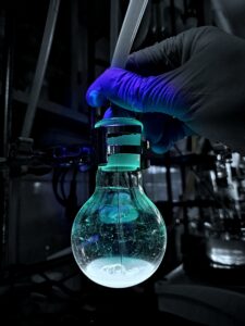
“Retro” Synthesis
Reductive aromatization of organic compounds in a retro look
Photo by Maciej Majdecki
|
|
|
Entry 17
|
Entry 18 |
|
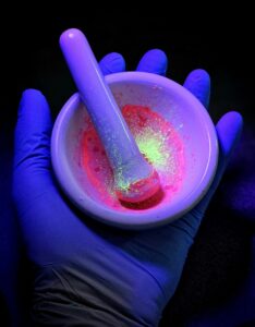
“Candy Mechanochemical Reaction”
Synthesis of a transition metal complex with a functionalized pyridine ligand
Photo by Maciej Majdecki
|
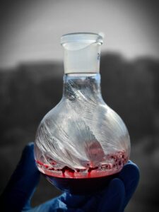
“Crystal Wings”
The effect of conducting the amidation reaction in imidazole
Photo by Maciej Majdecki
|
|
|
Entry 19
|
Entry 20 |
|
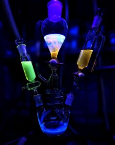
“Crazy Colourful Setup”
Color variation in synthetic setup
Photo by Maciej Majdecki
|
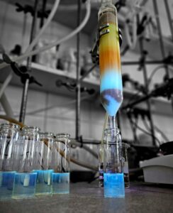
“Pacific Column Chromatography”
Chromatographic column exposed to UV radiation
Photo by Maciej Majdecki
|
|
|
Entry 21
|
Entry 22 |
|
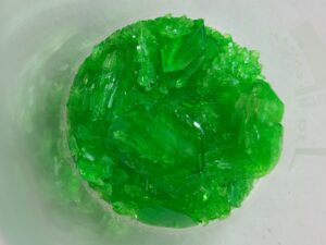
“Vibrant Green”
Crystals of potassium tris(oxalato)ferrate(III)
Photo by Helena Nogueira
|
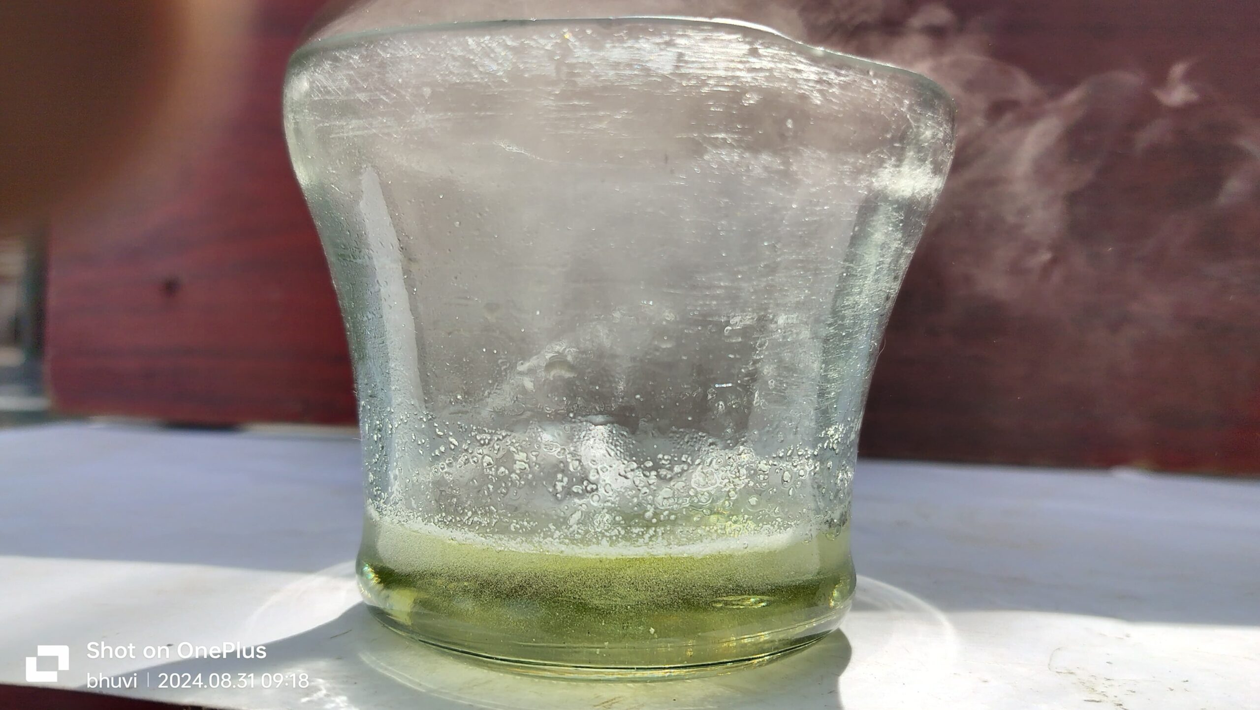
“Chemical reaction”
Aluminium reacts with hydrochloric acid to form aluminium chloride and hydrogen gas.
Photo by Gurusaran B.
|
|
|
Entry 23
|
Entry 24 |
|
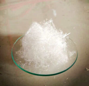
“Cotton candy”
Crystals of phthalic unhydride
Photo by Indhu Arunachalam
|
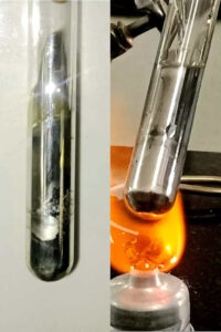
“Silver mirror 🪞”
Tollen’s reagent – It is a qualitative test which is used to distinguish between an aldehyde and a ketone
Photo by Indhu Arunachalam
|
|
|
Entry 25
|
Entry 26 |
|
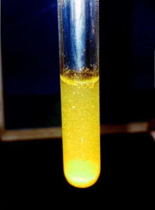
“Golden rain”
CGF – ‘chemical gold field ‘
Lead nitrate + potassium iodide to form lead iodide (yellow ppt)
Photo by Indhu Arunachalam
|
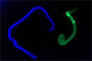
“Fluorescent anisakis: a worm of light”
Recently dead Anisakis simplex, the fairly common parasite of cold seas fish, emits a blue or green fluorescence, depending on the wavelength of excitation
Photo by Antonio D. Molina-Garcia
|
|
|




I love this
The pictures are so nice and it’s all about chemistry