Cellular organelles interact with one another to fulfill different functions within an organism. For example, interactions between mitochondria and lysosomes (membrane-enclosed organelles), such as mitophagy, participate in important cellular processes. Mitophagy is a selective form of autophagy (cellular recycling), in which defective or unnecessary mitochondria are isolated and fuse with lysosomes for degradation. Mitophagy regulates cell survival, cell proliferation, and cell death under stress conditions, and the dysregulation of mitophagy can cause various diseases. The typical mitophagy inducers are reactive oxygen species (ROS).
Methods for the real-time monitoring of interactions between mitochondria and lysosomes in cells, as well as ROS levels, could be useful to increase the understanding of how mitochondria-related diseases come about. They could also be important for monitoring the progression of diseases and optimizing treatment. Fluorescence imaging to trace mitophagy usually requires two dyes that label lysosomes and mitochondria separately with different emission colors. Developing single probes that can be used for the high-resolution visualization of the dynamic mitophagy process and evaluating cellular oxidative stress states is challenging.
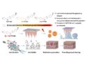
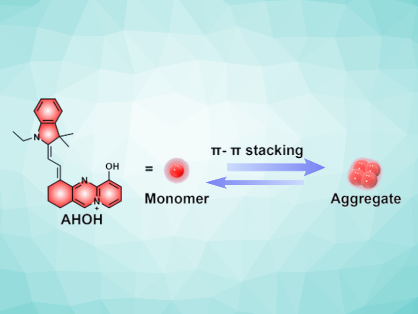
Hongbao Fang, Nanjing University, China, and Nanjing Normal University, China, Weijiang He, Yuncong Chen, Nanjing University and Nanchuang (Jiangsu) Institute of Chemistry and Health, Nanjing, China, and colleagues have designed and synthesized a ratiometric HClO probe called AHOH (pictured above), which can simultaneously stain lysosomes and mitochondria with red and green color, respectively (pictured below).

The chemical structure of AHOH is based on a merocyanine skeleton, which is nearly planar and readily self-assembles to form red-emitting nanoparticles, which accumulate in lysosomes via endocytosis. The monomeric form of AHOH tends to accumulate in mitochondria, is then oxidized by mitochondrial basal HClO, and shows green emission.
The probe could simultaneously monitor both mitochondria-lysosome interactions and oxidative stress through dual-channel high-resolution fluorescent imaging. AHOH could also be used in an imaging platform for evaluating drugs that regulate mitochondrial diseases.
- Aggregation‐based dual‐target probe for dual‐colour super‐resolution monitoring mitophagy and evaluating drugs regulating mitochondria,
Xiu‐Zhi Yang, Hongbao Fang, Shumeng Li, Chengyan Chu, Yunhua Zhang, Ying Yang, Weijiang He, Yuncong Chen, Zijian Guo,
Aggregate 2024.
https://doi.org/10.1002/agt2.641
![]()