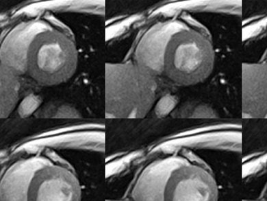
Jens Frahm and colleagues, Max Planck Institute for Biophysical Chemistry, Göttingen, Germany, have succeeded in significantly reducing the time required for recording cross-sectional images of our body by magnetic resonance imaging (MRI). The reduction of the image acquisition time to one fiftieth of a second (20 ms) makes it possible to obtain “live recordings” of moving joints and organs at previously inaccessible temporal resolution and without artefacts.
It relys on the already used FLASH (fast low angle shot) technique, but uses a radial encoding of the spatial information which renders the images insensitive to movements. The new mathematical reconstruction technique enables calculation of a meaningful image, in the most extreme case, out of just 5 % of the data required for a normal image.
Although these fast MRI measurements can be easily implemented on today’s MRI devices, the availability of sufficiently powerful computers for image reconstruction is something of a bottleneck: If, e.g., the heart is examined the computer system requires about 30 min at present to process one minute’s worth of film.
- Real-time MRI at a resolution of 20 ms
Martin Uecker, Shuo Zhang, Dirk Voit, Alexander Karaus, Klaus-Dietmar Merboldt, Jens Frahm
NMR in Biomed. 2010, 23.
DOI: 10.1002/nbm.1585 -
Related links:
[1] Live video of a beating heart
[2] Live video of movements during speech production
[3] Biomedizinische NMR Forschungs GmbH, Max Planck Institute for Biophysical Chemistry, Göttingen

