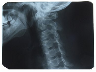
The destruction of myelin, an insulating sheet that enables neurons to correctly propagate electrical information, causes the development of severe neurological diseases, such as autoimmune encephalomyelitis. Imagining techniques able to quantify myelin in a noninvasive way are essential for the development and the validation of myelin-repair therapies.
Chunying Wu, Case Western Reserve University, Cleveland, USA, and colleagues developed a novel probe to monitor myelin by using the high resolution, nuclear imagining technique position emission tomography (PET). The use of the new compound [11C]MeDAS (11C-labeled N-methyl-4-4’-diamminostilbene), provided sensitive and specific images of myelin sheets undergoing destruction in the spinal cord of rats suffering from autoimmune encephalomyelitis. In addition, by using [11C]MeDAS they were able to quantify myelin-repair processes occurring in the spinal cord of diseased rats after treatment with hepatocyte growth factor, a protein which induces myelin regeneration.
[11C]MeDAS is, therefore, a promising tool to evaluate myelin-repair treatments.
- Longitudinal PET imaging for monitoring myelin repair in the spinal cord,
Chunying Wu, Junqing Zhu, Jonathan Baeslack, Anita Zaremba, Jordan Hecker, Janet Kraso, Paul M. Matthews, Robert H. Miller, Yanming Wang,
Ann. Neurol. 2013.
DOI: 10.1002/ana.23965


