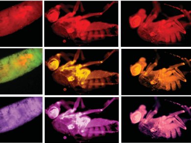
The fruit fly, Drosophila melanogaster, is one of the most prominent models to study developmental biology. Bioimaging of flies can be difficult — the handling of flies inside an NMR tube has been described as a very tricky job. Another option is optical imaging using fluorescent nanomaterials as contrast agents. These need to be introduced by intravenous/intramuscular injection or by gene translation, both of which can be difficult and time consuming.
Sabyasachi Sarkar and colleagues, Indian Institute of Technology, Kanpur, have developed a much simpler way to introduce fluorescent nanomaterials into fruit flies. They use water-soluble carbon nano-onions made from wood waste. The nano-onions are added to the fruit flies’ food and ingested orally by the flies. Just 4 ppm of nano-onions allowed the optical fluorescence microscopy imaging of all the stages of the fruit fly life cycle. The fluorescent fruit flies showed no toxic effects — upon withdrawal of the nano-onions from the food, they excreted the fluorescing material and continued to proliferate to the next generation, demonstrating a return to their normal lives.
This technique may be used in the easy, noninvasive image-based studies of other animals, and could ultimately be extended to human beings.
Image: © Wiley-VCH
- Carbon Nano-onions for Imaging the Life Cycle of Drosophila Melanogaster
M. Ghosh, S. K. Sonkar, M. Saxena, S. Sarkar,
Small 2011.
DOI: 10.1002/smll.201101158


