X-Ray Analysis
The determination of molecular structures by using X-ray analysis has allowed researchers to identify a huge number of chemical molecules. As a result, a vast amount of information on bond lengths and angles has been obtained. It has saved synthetic chemists a considerable amount of work because it is no longer necessary to perform very laborious total syntheses to prove the structure of a chemical compound.
A lesser known fact is that in some cases the total synthesis led to an erroneous result. As described by Dunitz [1], probably the most famous was the structure of patchouli alcohol, which was incorrectly assigned as (1) for more than 50 years [2]. The assignment was considered to be completed on the basis of the total synthesis but X-ray analysis gave structure (2) [3].
.gif)
Figure 1. The earlier proposed structure (1) and actual structure (2) of patchouli alcohol.
Disadvantages
Despite its advantages, X-ray diffraction also has drawbacks and limitations. Until now, one of the most important requirements is to have crystals of the compound, which then are examined. Another restriction is that it is impossible to study dissolved molecules. However, in solution, molecules can adopt various conformations unlike in the crystalline state, in which as a rule only one frozen conformation of the molecule exists. Interestingly, the conformation in the solid state is not necessarily the most stable one. For instance, [3.3]paracyclophane crystallizes in the trans conformation 3, whereas the cis conformation 4 is more stable (Fig. 2) [4].
.gif)
Figure 2. trans-[3.3]Paracyclophane 3 and cis-[3.3]paracyclophane 4.
Fluctuation X-Ray Scattering
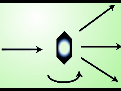
Recently, Zwart, Baylor College of Medicine, Houston, USA, and co-workers developed an extension of the X-ray diffraction method called fluctuation X-ray scattering. It allows one to determine the structure of proteins in solution much more accurately than multidimensional NMR techniques. The method proposed by Zwart‘s group [5, 6] involves laser irradiation of the sample with very high intensity X-ray pulses that have a very short lifetime of 10–15 s. Thus, this method allows one to observe the instantaneous conformation of the molecule because the pulse lasts less than a conformational transition. Unfortunately, the energy of the pulses is so high that after absorption the molecule falls apart. Hence, the method has also been called “scatter, diffract, and destroy” [7].
The method developed by Zwart’s group allows for computational analysis of data from a large number of scattering particles and determination of low-resolution structures of proteins and other macromolecules in solution with much greater accuracy than that of the determinations obtained so far by using less powerful synchrotron light sources. These limitations are especially consequential for proteins, which are difficult to study because of their size.
References
[1] J. D. Dunitz, X-ray Analysis and the Structure of Organic Molecules, Cornell University Press, Ithaca, 1979, pp. 325, 337, 363.
(For a newer edition see, J. D. Dunitz, X-ray Analysis and the Structure of Organic Molecules, Second Edition, Verlag Helvetica Chimica Acta, Zürich, Switzerland, 1995. ISBN: 9783906390390)
[2] G. Buchi, William D. Macleod, J. Am. Chem. Soc. 1962, 84, 3205–3206. DOI: 10.1021/ja00875a047
[3] M. Dobler, J. D. Dunitz, B. Gubler, H. P. Weber, G. Büchi, J. Padilla, Proc. Chem. Soc. 1963, 383. DOI: 10.1039/PS9630000357
[4] S. Szymański, H. Dodziuk, M. Pietrzak, J. Jaźwiński, T. B. Demissie, H. Hopf, J. Phys. Org. Chem. 2013, 26, 596. DOI: 10.1002/poc.3137
[5] Gang Chen, Peter H. Zwart, Dongsheng Li, Phys. Rev. Lett. 2013, 110, 195501. DOI: 10.1103/PhysRevLett.110.195501
[6] Haiguang Liu, Billy K. Poon, Dilano K. Saldin, John C. H. Spence, Peter H. Zwart, Acta Cryst. A 2013, 69, 365–373. DOI: 10.1107/S0108767313006016
[7] David Bradley, Diffract and destroy: Fluctuation X-ray scattering, SpectroscopyNow.com, June 1, 2013. Link
More on the International Yeat of Crystallography (IYCr2014)


