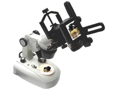
All major manufacturers of microscopes also supply cameras for imaging objects. As these are expensive, Ekkehard Diemann, University of Bielefeld, Germany, describes low-budget solutions for imaging objects by using a simple compact digital camera, a simple microscope, and a tripod. The results are excellent for illustrating experimental protocols, laboratory notebooks, or experimental instructions.
In the simplest case, the camera is positioned in front of the eyepiece of the microscope, which has already been focused on the object. The camera can be positioned with a tripod, or, much safer, it can be attached directly to the tube of the microscope (pictured). Special sockets for this purpose are available as photo equipment.
If the camera allows it, one can make an exposure bracketing, e.g., a normally exposed image and in addition one image with one aperture stop over-exposed and another one under-exposed. This procedure can be repeated with slightly modified lighting, if details are obscured by shadows. Exposure bracketing allows the extension of the otherwise low dynamic range of the image sensor, i.e., details in dark and light areas of the images can be made more visible, than they would be with a single shot.
The last step is the image processing, e.g., when only parts of the image are of interest, color casts must be corrected, or contrast, brightness or black point should be optimized. For these tasks, numerous free programs are available.
In the second part of the article, Diemann introduces scanography, a method to reproduce 3D objects by using a flatbed scanner. The color fidelity is very good and the depth of field is surprisingly high. The illumination provided by the revolving LED strip is usually optimal, shadows rarely form or can be suppressed by a scattering foil, as they are used to protect smartphone displays, on the glass plate.
- Digitalfotografie in der Chemie,
Ekkehard Diemann,
Chem. Unserer Zeit 2013, 47, 376–380.
DOI: 10.1002/ciuz.201300630


