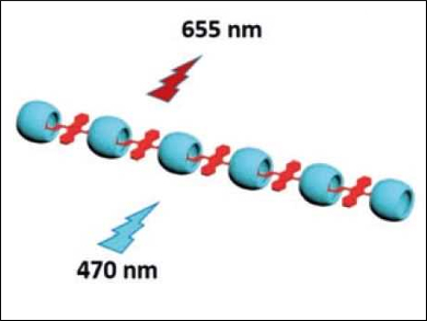
Supramolecular assemblies containing dyes can be used for cell imaging, even targeted to specific cell organelles. Yu Liu, Nankai University and Collaborative Innovation Center of Chemical Science and Engineering, both Tianjin, China, and colleagues have developed supramolecular assemblies for lysosome-targeted cell imaging. The lysosome is an organelle composed of spherical vesicles which contain enzymes.
The team used the organic dye 4,4’‐anthracene‐9,10‐diylbis(ethene‐2,1‐diyl)bis(1‐ethylpyridin‐1‐ium) bromide (ENDT), which has a very weak fluorescence emission at 625 nm. When ENDT forms a complex with cucurbit[8]uril, the complex assembles into nanorods (pictured) with a near‐infrared (NIR) fluorescence emission at 655 nm. This change in the emission spectrum is caused by the aggregation of the dye molecules. Further complexation with a modified sulfonatocalix[4]arene leads to another fluorescence enhancement.
NIR light causes little damage to biological samples and allows for deep tissue penetration. The researchers showed that the synthesized assemblies can be used as targeted imaging agents for lysosome in living cells. This could be used for the detection of lysosome activities and, ultimately, in medical diagnosis.
- Supramolecular Assemblies with Near-Infrared Emission Mediated in Two Stages by Cucurbituril and Amphiphilic Calixarene for Lysosome-Targeted Cell Imaging,
Xu-Man Chen, Yong Chen, Qilin Yu, Bo-Han Gu, Yu Liu,
Angew. Chem. Int. Ed. 2018.
https://doi.org/10.1002/anie.201807373


