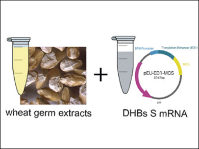
Obtaining three-dimensional representations of complex membrane protein assemblies is important for health-related research, where this information can be used to guide drug development. However, structural determination often faces two problems: how to reproduce protein assembly in a native form in vitro, and how to analyze its 3D structure in this environment. Cell-free protein expression and solid-state NMR spectroscopy are promising approaches in this context. To date, they have been largely incompatible with obtainable sample amounts. However, recent NMR techniques, such as magic angle spinning (MAS) solid‐state NMR at >100 kHz, have reduced the required amount by nearly a factor of hundred.
Anja Böckmann, Université de Lyon, France, Beat H. Meier, Swiss Federal Institute of Technology (ETH) Zurich, Switzerland, and colleagues have demonstrated that subviral particles of the duck hepatitis B virus (DHBV) made from an integral membrane protein can be produced and spontaneously self-assemble in a wheat‐germ cell-free protein expression. Duck hepatitis B is an important model for the human virus.
MAS solid-state NMR spectra of as little as 0.5 mg of these assemblies can be recorded with sufficient resolution and sensitivity to initiate structural studies. These results open the way to structural studies of viral envelope protein assemblies that are central in infection, and can also be applied to other complex membrane proteins.
- Structural Studies of Self-Assembled Subviral Particles: Combining Cell-Free Expression with 110 kHz MAS NMR Spectroscopy,
Guillaume David, Marie-Laure Fogeron, Maarten Schledorn, Roland Montserret, Uta Haselmann, Susanne Penzel, Aurélie Badillo, Lauriane Lecoq, Patrice André, Michael Nassal, Ralf Bartenschlager, Beat H. Meier, Anja Böckmann,
Angew. Chem. Int. Ed. 2018.
https://doi.org/10.1002/anie.201712091


