Organ-Targeted Gold Catalysis
A gold catalyst can be delivered to a target organ in a higher organism where it performs a chemical transformation visualized by bioimaging. This intriguing approach has been introduced by a Japanese team of scientists in the journal Angewandte Chemie. It could make organometallic catalysis applicable for therapy or diagnostics.
How is a therapeutically useful catalyst guided into its target tissue to synthesize bioactive molecules and drugs in higher levels? How can its activity be visualized there? Noninvasive targeting for therapy, biological sensing, and imaging has become one of the most active biomedical research areas. Katsunori Tanaka and his colleagues at RIKEN, Waseda University, and JST-PRESTO, Japan, and Kazan Federal University, Russia, are especially interested in biocompatible metal complexes and, among them, gold catalysts to perform synthetic transformations in a target tissue. The challenge, however, is to bring the gold specifically to its target organ and to establish a visualization scheme to monitor the ongoing biochemical transformation.
Glycoalbumins Direct Metal Complexes
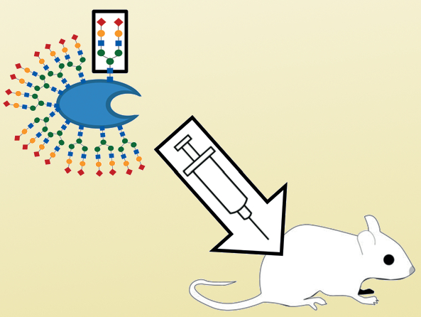
Gold ions can be conjugated to a hydrophobic protein ligand, and this complex can be bound to albumin, an abundant water-soluble protein. The albumin is then furnished with sugar-type molecules, the glycans, which carry the chemical groups responsible for glycoalbumin accumulation in a target organ: “This work explores the adaptation and usage of organ-targeting glycans as biologically compatible metal carriers,” the scientists wrote. Thus, the glycoalbumin can deliver the biocompatible metal catalyst, namely the gold complex. Intriguingly, this gold complex efficiently acts as an organometallic catalyst that can perform the reaction between biologically relevant molecules and organic substrates, which means it could be a relevant drug or diagnostic compound.
The scientists used the gold complex to bind a fluorescent dye to certain surface proteins present in the target tissue, which was either the liver or the intestine. To visualize the reaction, they performed fluorescent imaging of the whole living mouse. Within two hours after the injection of the catalyst and the substrate (the functionalized fluorescent dye) into the blood circuit, strong fluorescence in the two organs demonstrated successful in vivo gold catalysis. Thus, a catalytically active gold complex was sent and delivered to a target organ within a short time and without the laborious development of antibodies. As an outlook the scientists envisage biomedical applications, especially for metal catalysts with their unique reactivities: “Example therapies may include uncaging of active, cancer therapeutic enzymes selectively at tumor sites or […] reactions to produce active drug molecules at targeted organs,” they wrote.
- In Vivo Gold Complex Catalysis within Live Mice,
Kazuki Tsubokura, Kenward K. H. Vong, Ambara R. Pradipta, Akihiro Ogura, Sayaka Urano, Tsuyoshi Tahara, Satoshi Nozaki, Hirotaka Onoe, Yoichi Nakao, Regina Sibgatullina, Almira Kurbangalieva, Yasuyoshi Watanabe, Katsunori Tanaka,
Angew. Chem. Int. Ed. 2017.
DOI: 10.1002/anie.201610273


