A blood vessel is crucial for circulatory diseases and treatments, the biological evaluation of drug diffusion to target tissues, the penetration of cancer cells or pathogens, and the control of blood pressure. It is generally composed of three distinct layers: the intima, an inner single layer of endothelial cells (ECs); the media, medium layers of smooth muscle cells (SMCs); and the adventitia, an outer layer of fibroblast cells.
Nitric oxide is a lipophilic, highly diffusible, and short-lived physiological messenger. Quantitative, kinetic, and spatial analyses of the extracellular delivery of NO molecules from the EC layer to the SMC layers upon drug stimulation are crucial for pharmaceutical and biomedical evaluations of hypertension and diabetes.
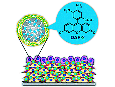
Mitsuru Akashi and his team, Osaka University, Japan, demonstrate a three-dimensional analysis of NO diffusion in a blood vessel in response to drug stimulation by using 3D artery models including sensor particles (see picture). The quantity and diffusion distance of NO in the five-layered models were almost the same as those for the in vivo blood vessel response. The method does not require special instruments or techniques and is convenient and effective for drug screening. Thus it has possibility to be a solution of general animal experiments.
- Quantitative 3D Analysis of Nitric Oxide Diffusion in a 3D Artery Model Using Sensor Particles,
Michya Matsusaki, Suzuka Amemori, Koji Kadowaki, Mitsuru Akashi,
Angew. Chem. Int. Ed. 2011, 50, 7557-7561.
DOI: 10.1002/anie.201008204


