Understanding the processes inside the nucleus of a cell could lead to greater comprehension of genetics and the factors that regulate expression. Proteins and dyes used to track activity aren’t easy to use with microscopy techniques due to their weak signals and tendency to photobleach.
Min-Feng Yu and co-workers, University of Illinois, USA, have shown that streptavidin-conjugated quantum dots (QDs) with overall diameter of ≈15–20 nm offer the advantages of small size, bright fluorescence for easy tracking, and excellent stability in light.
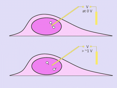
The QDs were delivered to the nucleus by a nanoneedle made from a boron nitride nanotube coated with gold. This was loaded with the QDs. An electrical charge was applied to release the QDs at the desired location and moment.
This technique provides a level of control not achievable by other molecular delivery methods, which involve gradual diffusion throughout the cell and into the nucleus.
- Electrochemically Controlled Deconjugation and Delivery of Single Quantum Dots into the Nucleus of Living Cells
K. Yum, N. Wang, M.-F. Yu,
Small 2010, 6 (19), 2109–2113.
DOI: 10.1002/smll.201000855


